
Behind every image is a patient.
Behind every patient is a care team trusted with getting it right.
Designed with your everyday in mind.
ASE Scientific Sessions | June 26-28 | Booth #503
Product Overview
Clarity you can stand behind.
Vivid™ Pioneer delivers extraordinary imaging, AI-powered automation, and one-click optimization to support consistent results, reduce variability, and streamline demanding workflows.1
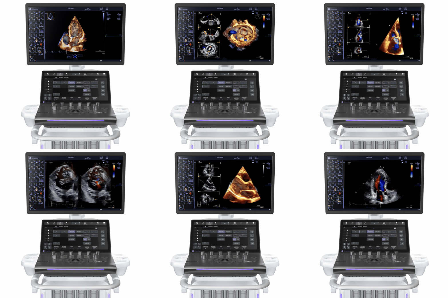

Sharper images
Easily identify small anatomical structures, such as valve leaflets

Precise images—first scan
Clearly see delineation of the myocardial wall from the first scan out of the box

Conquer demanding workflows
Streamline measurements and quantification with AI and automated tools

Light, highly portable
Bring ultra-premium imaging effortlessly into more and tighter places
Solutions designed to fit your workflow,
not the other way around.
Optison™
(Perflutren Protein Type A Microspheres Injectable Suspension, USP)
Ultrasound contrast enhancement that supports diagnostic cardiac imaging when standard image quality is suboptimal, enabling confident clinical decisions without adding complexity to existing workflows.
See important safety information below.

Vivid™ Ultra Edition*
High‑quality cardiovascular ultrasound imaging at the foundation, delivering clarity and reproducibility across echo labs and shared services.
*Ultra Edition is not a product name; it refers to the 2022 release of the Vivid portfolio.

Vscan Air™ SL with Caption AI™
A portable, handheld ultrasound that guides users to capture diagnostic quality echo images at the point of care.

ViewPoint EchoPilot™
A cloud based AI driven solution that automatically generates precise measurements and delivers a preview of your routine echocardiogram report in just seconds.

ViewPoint™ with EchoPAC™
A comprehensive reporting and image management solution designed to help clinicians focus more on patient care. With EchoPAC, create the workflow that works for you and enhance cardiac patient care beyond image acquisition.

Centricity™ Cardio Enterprise™
Streamline cardiology imaging and workflows across service lines bringing together images, measurements, and reports to enhance care continuity and support faster, more confident clinical decisions

Drug (Optison (Perflutren Protein-Type A Microspheres Injectable Suspension, USP) and Device (Vivid ) are NOT Combination Products as classified under 21 CFR 3.2(e). Both Drug & Device were approved/cleared independent of the other by the FDA. While the products can be used in conjunction with each other, Drug does not require the use of this specific Device, nor does Device require the use of this specific Drug.
Echo Education Hub
June 26–28
Learn how to acquire, analyze, and interpret advanced echo data using AI, strain, and 4D imaging in a hands-on, case-based environment.
Presenters

Christos Mihos, DO
Director, Echocardiography & Non-Invasive Cardiovascular Laboratory, Division of Cardiology Mount Sinai Medical Center | Miami, FL

Arash Sabati, MD, FACC
Director of Heart Center Research, Phoenix Children’s Hospital | Phoenix, AZ

Jennifer Liu, MD, FASE
Chief of Cardiology, Memorial Sloan Kettering Cancer Center | New York , NY

Salima Qamruddin, MD, MPH, FASE
Technical Director Echocardiography Laboratory, OchsnerMedical Center | New Orleans, LA

Renuka Jain, MD, FACC
Director of Echocardiography, Aurora St. Luke s Medical Center | Milwaukee, WI

Kiran Belani, MD, FASE, FACC
Assistant Professor of Anesthesiology and Cardiac Surgery, Northwestern Medicine | Chicago, IL

Jessica Stout, BS, ACS, RDCS, FASE
Director of Non-Invasive Cardiology, Beth Israel Deaconess Medical Center | Boston, MA

Pamela R. Burgess, BS, ACS, RDCS, RDMS, RVT, FASE
Cardiac Sonographer Educator,
Atrium Health Wake Forest Baptist | Winston-Salem, NC
Agenda
Friday, June 26
1:00 P.M. – 2:00 P.M. | From Pediatrics to Adults: Practical Applications of Optison-Enhanced Ultrasound
Join us for an in-depth educational program on the use of Optison to optimize ultrasound imaging across patient populations. This session will begin with a pediatric-focused presentation covering key case examples, administration techniques, and dosing considerations in pediatric imaging. The second presentation will transition to adult applications, showcasing ultrasound images enhanced with Optison and discussing how contrast optimization supports image quality and diagnostic in adult patients.
Presented by: Arash Sabati, MD, FACC and Pamela Burgess ACS, BS, RDCS, RDMS. RVT, FASE
2:15 P.M. – 3:15 P.M. | AFI Strain in Cardio-Oncology
• Understand how cancer therapy impacts cardiac function and why strain detects early injury
• Use GLS for early detection assessment and monitoring of cardiotoxicity over time
• Recognize meaningful strain changes and what they indicate clinically
• Apply strain insights to guide real-world clinical decisions using EchoPAC
Presented by: Jennifer Liu MD, FASE
3:30 P.M. – 4:15 P.M. | The 4th Dimension: Optimizing 4D Images and 4D Quantification of the LV and LA
• Learn how to optimize 4D Volumes
• Understand the relationships of image resolution, frame rate and volume size in 4D
• Discover how to quantify 4D Volumes of the Left Ventricle and Left Atrium
• Learn how to navigate a 4D Volume
• Get hands-on learning using 4D images to quantify the LA and LV in 4D
Presented by: Jessica Stout BS, ACS, RDCS, FASE
Saturday, June 27
10:15 A.M. – 11:00 A.M. | Intro to Strain Imaging: Bringing AFI into Your Echo Lab
• Introduction myocardial strain measurement and why it matters clinically
• Explore how artificial intelligence has enhanced strain analysis
• Apply concepts through real world patient case studies
• Learn proper measurement and editing techniques while avoiding common pitfalls
• Interpret myocardial strain results at both regional and global levels
Presented by: Christos Mihos, DO
11:15 A.M. – 12:00 P.M. | Artificial Intelligence in Echo: Transform Your Efficiency
• Understand the development and critical role of AI and machine learning in echo
• Explore current applications of AI in echo disease detection, automated measurements, and echo reporting
• Identify technologies that enhance efficiency and consistency in the echo lab
• Apply AI using EchoPAC for image analysis, measurement editing, and clinical interpretation
Presented by: Salima Qamruddin MD, MPH, FASE
1:30 P.M. – 3:00 P.M. | ASE Sponsored – 3D Pediatric Learning Lab
ASE session led by: Pei-Ni Jone, MD,FACC,FAHA,FASE; Nee Scze Khoo, MBChB,FRACP; Benjamin Goot, MD,
FASE, Lily Lin, MD
3:30 P.M. – 4:30 P.M. | 4D TEE of the Mitral Valve in Structural Imaging & Procedural Guidance
• Learn the importance of 4D Echo in Structural Heart Procedures
• Understand anatomical orientations and where 4D tools are used in Mitral procedures
• Discover MPRs (Flexi-Slice) and how to align views for MV procedures
• Get hands on learning using flexi-slice from 4D data sets on EchoPac
Presented by: Renuka Jain MD, FACC
Sunday, June 28
9:30 A.M. – 10:30 A.M. | Left Atrial Appendage Closures: How Can 4D and AI Elevate Your Program
• Understand the LAAO workflow and the role of imaging at each stage
• Leverage 4D imaging for procedural guidance and decision-making
• Explore AI tools that support LAAO workflow efficiency
• Interpret LAA anatomy using 4D TEE and 4D ICE
• Apply key measurements and emerging technologies for LAAO closure
• Work with 4D datasets using multiplanar reconstruction (FlexiSlice)
Presented by: Kiran Belani, MD, FASE, FACC
11:00 A.M. – 12:00 P.M. | 4D TEE of the Tricuspid Valve in Structural Imaging and Procedural Guidance
• Learn the importance of 4D Echo in Structural Heart Procedures
• Understand anatomical orientations and where 4D tools are used in Tricuspid procedures
• Discover MPRs (Flexi-Slice) and how to align views for TV procedures
• Get hands on learning using flexi-slice from 4D data sets on EchoPac
Presented by: Renuka Jain MD, FACC
1:00 P.M. – 2:00 P.M. | Left Atrial Strain Helping Us With HFpEF and Diastolic Function
• Understand the clinical value of LA strain
• Interpret the phases of LA strain and their significance
• Apply LA strain in assessing HFpEF and diastolic dysfunction
• Follow guideline-based approaches to measurement and interpretation
• Confidently measure LA strain and recognize common pitfalls
Presented by: Christos Mihos, DO
Science and Tech Theater
Unlocking New Possibilities in Echo
Saturday, June 27 | 12:30 P.M. – 1:30 P.M.
Includes catered lunch. Popular event arrive early to secure your seat.
Artificial intelligence, adaptive imaging, enhanced color flow, ergonomic design, LAA assessment, and real-time 4D ICE in clinical practice. Join expert faculty to explore how these innovations revolutionize imaging, improve efficiency, and support more confident clinical decisions.
Panel

Scott Chadderdon, MD
Cardiologist, Assoc. Prof. of Medicine, Oregon Health and Science University (OHSU) | Portland, OR

Renuka Jain, MD, FACC
Director of Echocardiography, Aurora St. Luke's Medical Center | Milwaukee, WI

Ashlee Davis, ACS, RDCS, RCCS
Chief Technologist, Duke University Hospital | Durham, NC

Denise Ignatowski, BS, RDCS, FASE
Echo Lab Manager, Aurora St Luke s Medical Center | Milwaukee, WI

Jacob Dewees, RDCS
Clinical Luminary Leader, GE HealthCare
Real-World Evidence Case Study from Piedmont Healthcare Cardiovascular Imaging | Meet the Authors
Explore a real-world evidence case study from Piedmont Healthcare highlighting the safety profile of Optison (Perflutren Protein-Type A Microspheres Injectable Suspension, USP) in cardiovascular imaging. Connect with Jackie Helm, ACS, RDCS, RVT, FASE, and Alecia Bradley, BSDMS, RDCS, RVT, RDMS—experienced cardiovascular imaging specialists and co-authors of this important case study. They share expert insights from their research, including practical considerations, safety observations, and the role of contrast-enhanced imaging in advancing cardiac diagnostics.
Gain valuable perspective on how real-world data and innovative imaging techniques are helping to improve outcomes in cardiovascular care.
Authors

Jackie Helm, ACS, RDCS, RVT, FASE
Piedmont HealthCare | Atlanta, GA

Alecia Bradley, BSDMS, RDCS, RVT, RDMS
Piedmont HealthCare | Atlanta, GA
Echo Discovery Zone
Saturday, June 27 | 1:30 P.M. – 3:30 P.M.
Optison Case Studies Demos and IV Insertion Training
Saturday, June 27 | 1:30 P.M. – 3:30 P.M.
Valvular and Structural 4D TEE Imaging
Sunday, June 28 | 9:00 A.M. – 12:00 P.M.
4D TTE Acquisition Optimization
At GE HealthCare, we re at the heart of it all. That s because we re designing tools tailored to your every day that work just as hard as you do.
See you at ASE 2026!
Important Safety Information
WARNING: SERIOUS CARDIOPULMONARY REACTIONS
Serious cardiopulmonary reactions, including fatalities, have occured uncommonly during or following perflutren-containing microscopic administration.
Most serious reactions occur within 30 minutes of administration
• Assess all patients for the presence of any condition that precludes OPTISION administration
• Always have resuscitation equiment and trained personnel readily available
OPTISON (Perflutren Protein-Type A Microspheres Injectable Suspension, USP)
INDICATIONS:
OPTISON is an ultrasound contrast agent indicated for use in adult and pediatric patients with suboptimal echocardiogram to opacify to the left ventricle to improve the delineation of the left ventricle endocardial borders.
CONTRAINDICATIONS:
OPTISON is contraindicated in patients with known or suspected hypersensitivity to perflutren or albumin.
WARNINGS AND PRECAUTIONS:
- Serious cardiopulmonary reactions including fatalities have occurred uncommonly during or shortly following perflutren-containing microsphere administration, typically within 30 minutes of administration. The risk for these reactions may be increased among patients with unstable cardiopulmonary conditions (acute myocardial infarction, acute coronary artery syndromes, worsening or unstable congestive heart failure, or serious ventricular arrhythmias).
- Serious anaphylactic reactions have been observed during or shortly following perflutren-containing microsphere administration, including shock, hypersensitivity, bronchospasm, throat tightness, angioedema, edema (pharyngeal, palatal, mouth, peripheral, localized), swelling (face, eye, lip, tongue, upper airway), facial hypoesthesia, rash, urticaria, pruritus, flushing, and erythema have occurred in patients with no prior exposure to perflutren-containing microsphere products.
- When administering OPTISON to patients with a cardiac shunt, microspheres can bypass filtering of the lungs and enter the arterial circulation. Assess patients with shunts for embolic phenomena following OPTISON administration. OPTISON is only for intravenous administration; do not administer OPTISON by intra-arterial injection.
- High ultrasound mechanical index values may cause microsphere rupture and lead to ventricular arrhythmias. Additionally, end-systolic triggering with high mechanical indices has been reported to cause ventricular arrhythmias. OPTISON is not recommended for use at mechanical indices greater than 0.8.
Adverse Reactions
Common adverse reactions (incidence > 0.5%) were: headache, nausea and/or vomiting, warm sensation or flushing, dizziness, dysgeusia, chills or fever, flu-like symptoms, malaise/weakness/fatigue, chest pain, dyspnea, injection site discomfort, and erythema. Cardiac arrests and other serious but nonfatal adverse reactions were uncommonly reported in post-approval use. Reports also identified neurologic reactions (loss of consciousness or convulsions) as well as anaphylactoid reactions. Overall, the safety profile observed in pediatric patients from the clinical study was consistent with the safety profile in adult patients.
Use in Specific Populations
Pregnancy and Lactation:
There are no data with OPTISON use in pregnant woman to inform any drug-associated risks. There are no data on the presence of perflutren protein-type A microspheres in human milk, the effects on the breastfed infant or the effects on milk production.
Pediatric Use
Safety and efficacy of OPTISON in pediatric patient is supported by evidence from adequate and well-controlled studies in adults and additional efficacy and safety data from a clinical study in 37 pediatric patients aged 9-17 years.
Geriatric Use
No overall differences in safety or effectiveness were observed in patients 65 years and over but a greater sensitivity to OPTISON in older individuals cannot be ruled out.
Please see the full Prescribing Information, including Boxed Warning for additional important safety information.
To report SUSPECTED ADVERSE REACTIONS, contact GE HealthCare at 800 654 0118 (option 2 then option 1) or by email at [email protected] or FDA at 800 FDA 1088 or www.fda.gov/medwatch
Reference
- The Role of AI in Streamlining Echocardiography Quantification White Paper, Kristin McLeod and Jurica Sprem, Senior Data Scientists at GE HealthCare. JB20789XX. A copy of this file can be available upon request.


Privacy policy | Terms and Conditions | Email Preferences | Contact us
© 2026 GE HealthCare.
Vivid, Vscan Air, Caption AI, Viewpoint, EchoPilot, Centricity, Optison, EchoPAC are trademark of GE HealthCare.
GE is a trademark of General Electric Company used under trademark license.
June 2026 | JB13890US