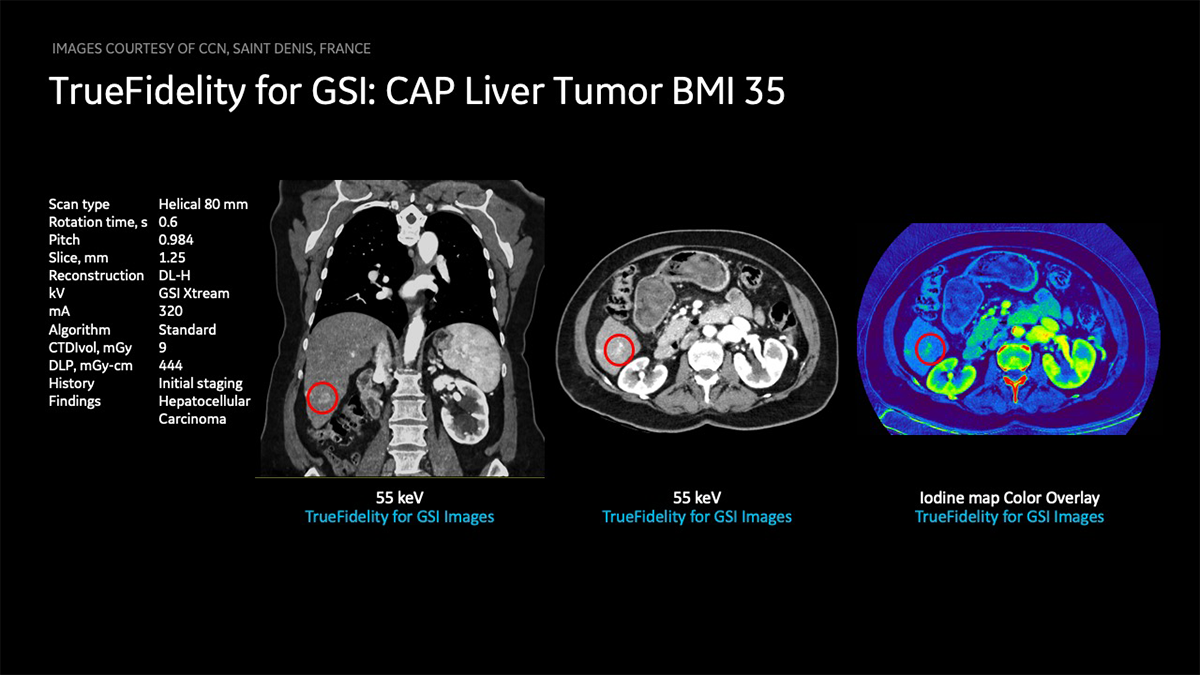
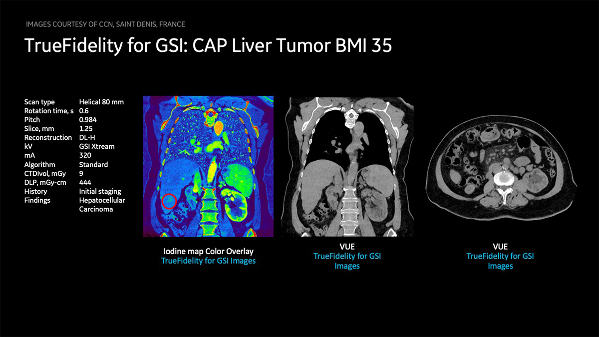
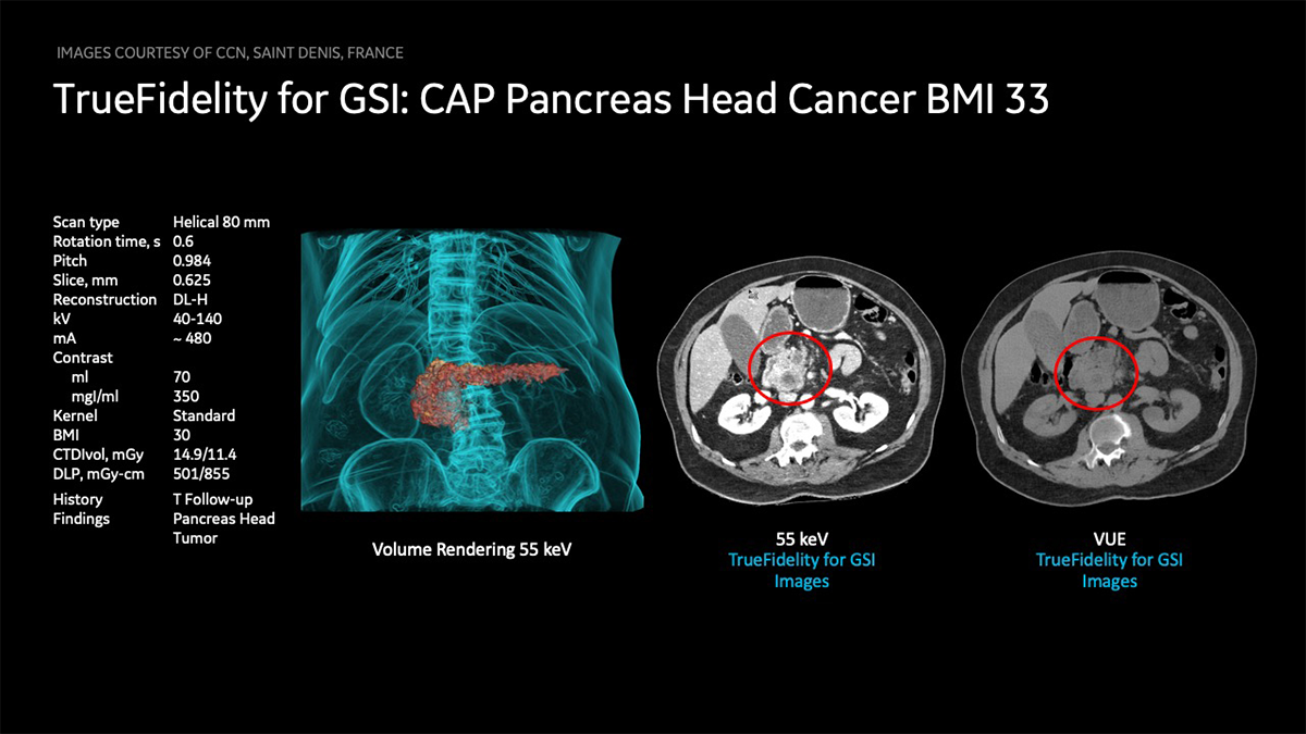
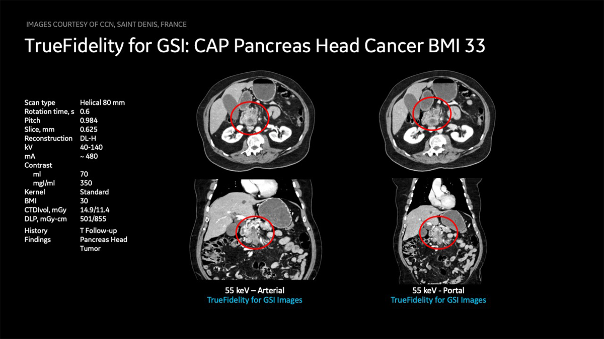
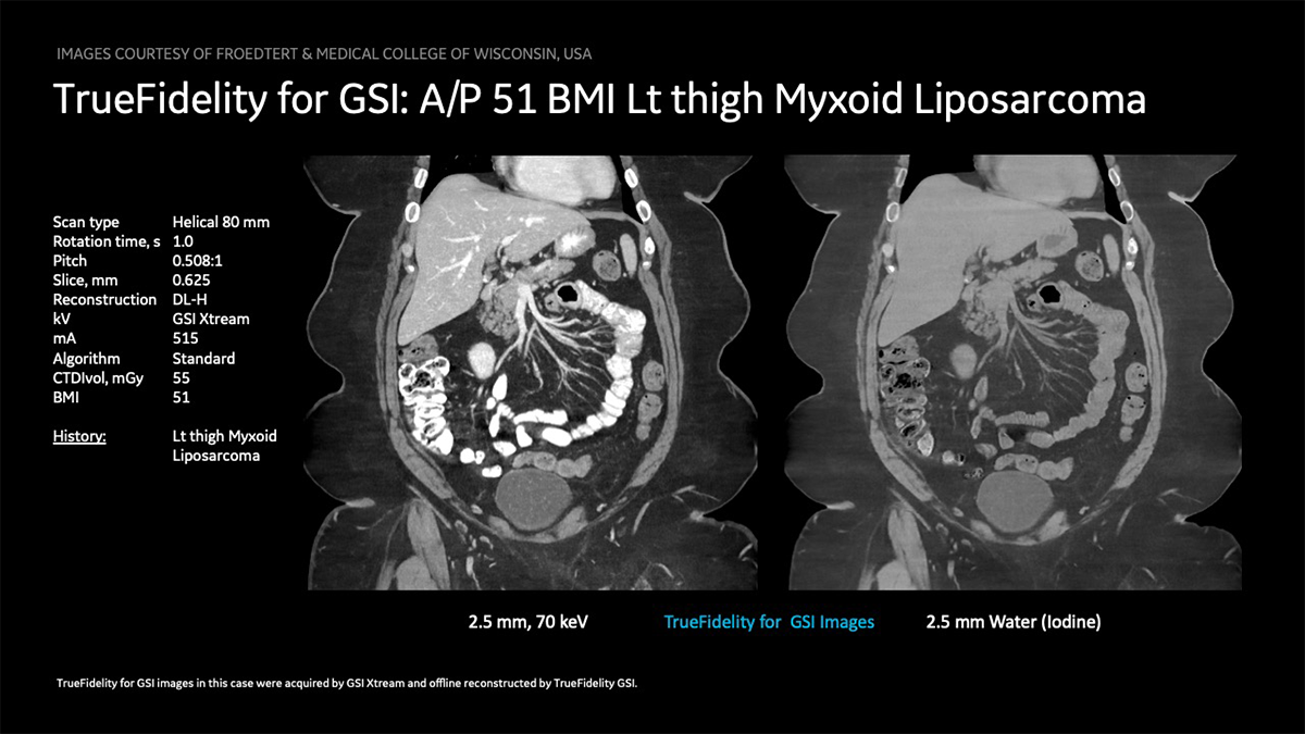
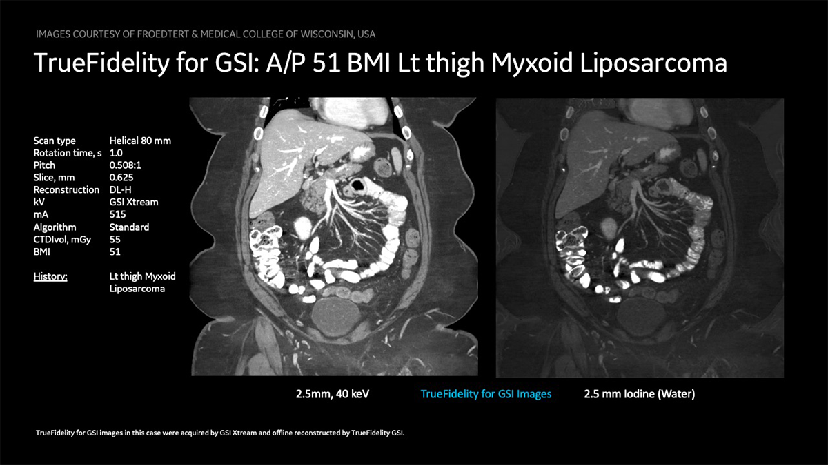
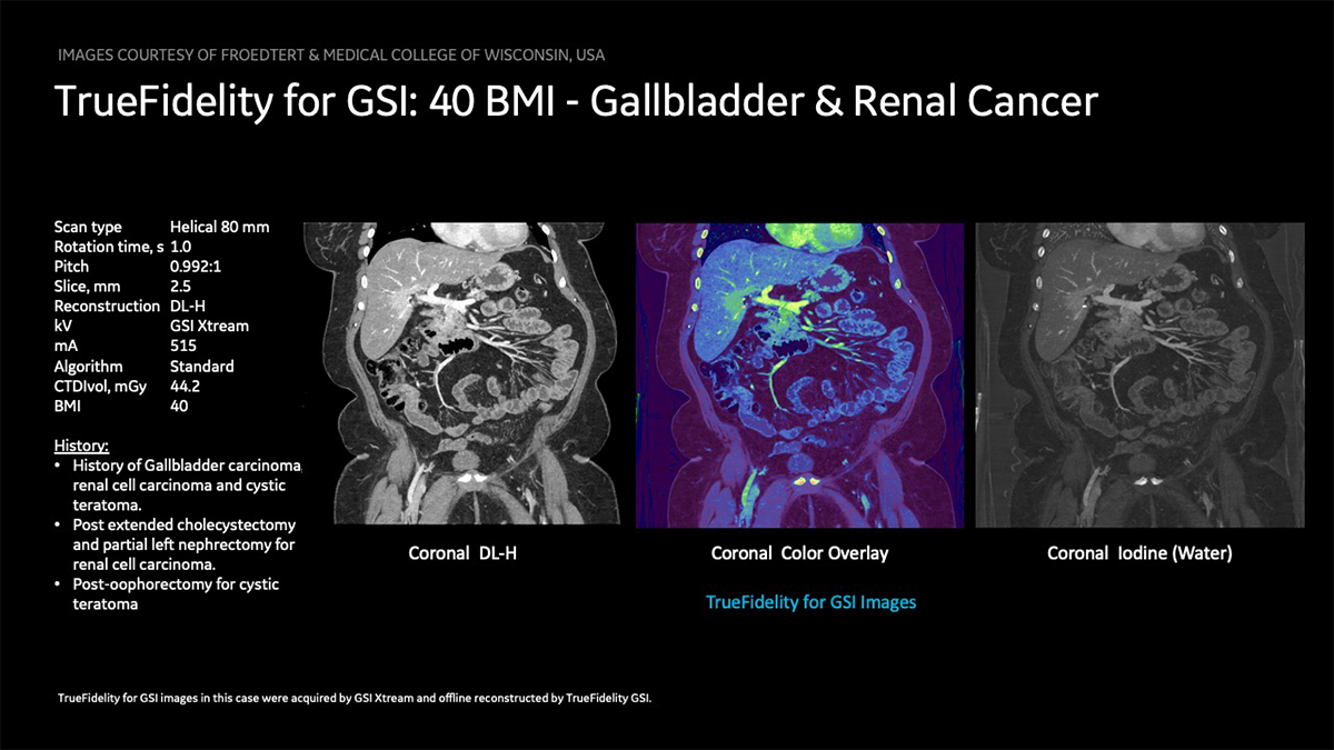
TrueFidelity for GSI has the potential to improve the image quality of GSI, especially of low-energy monochromatic images (40–70 keV), iodine images and VUE images which are critical for oncology imaging:
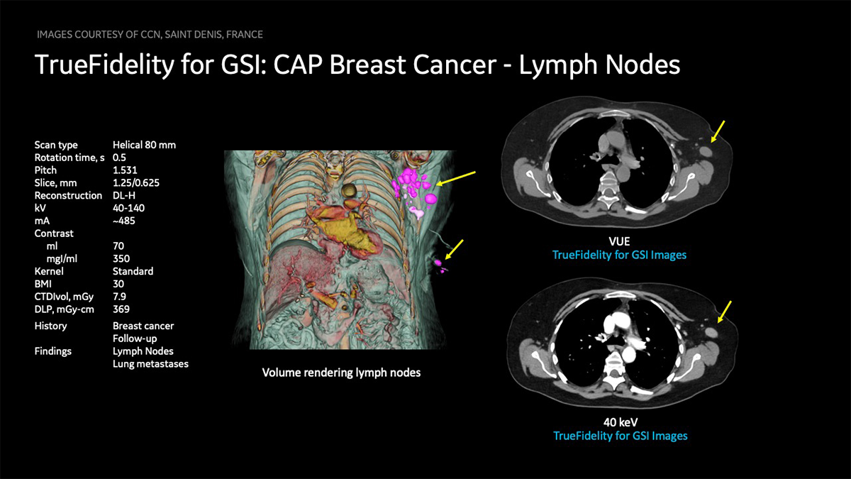
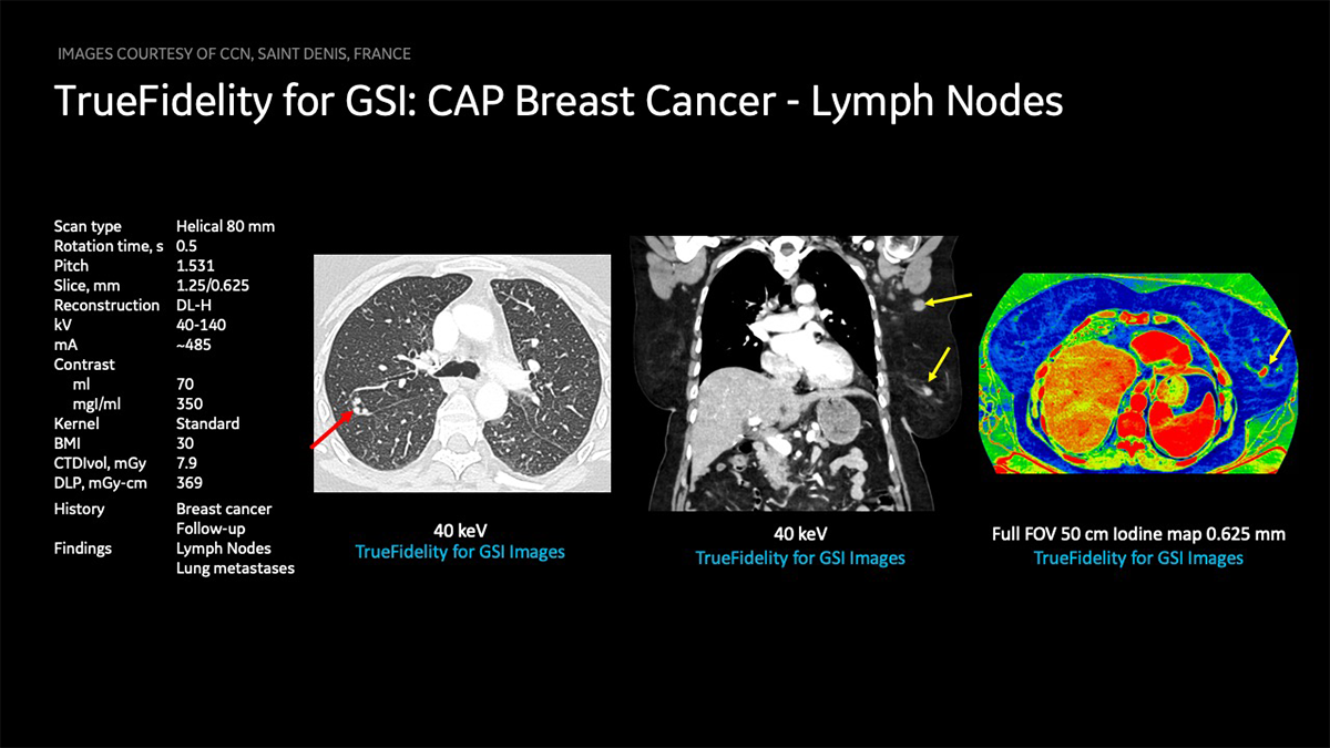
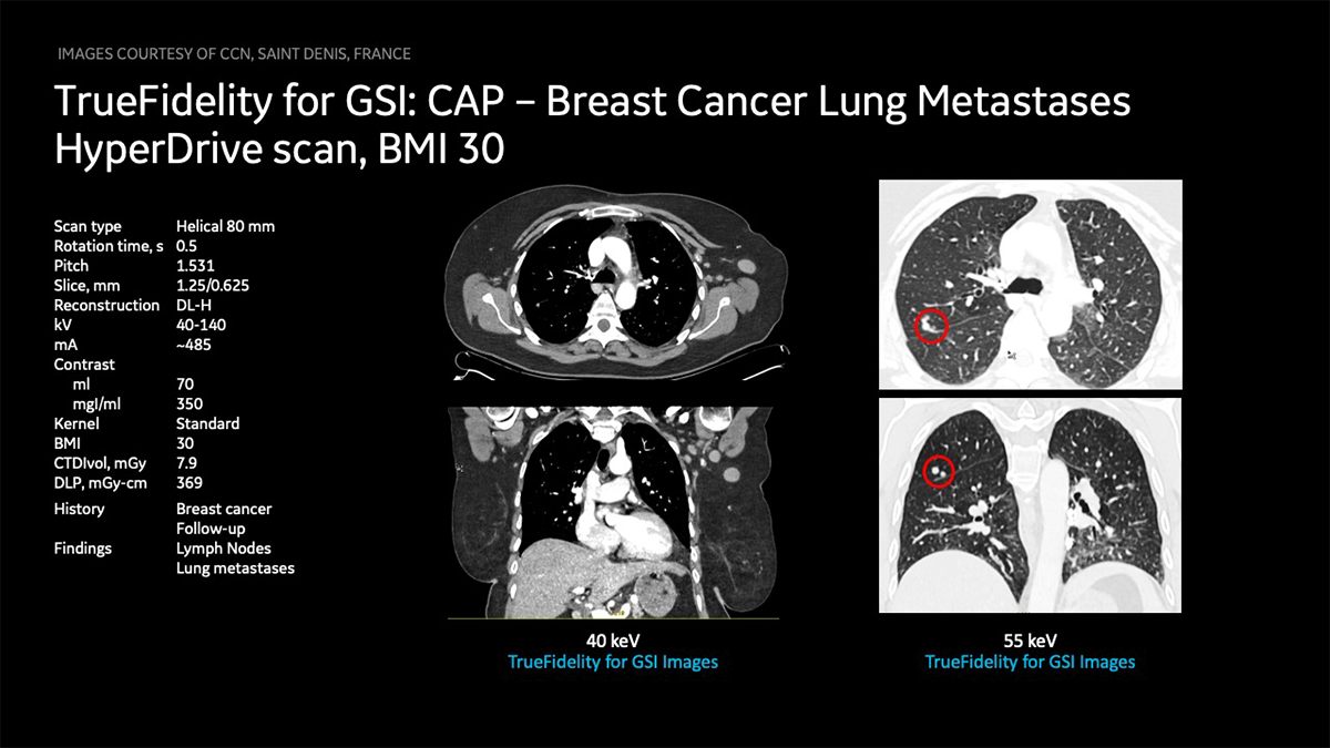
• The use of low-energy monochromatic images in oncologic CT can improve lesion detection by improving the contrast between a hypervascular lesion, a hypovascular lesion, and normally enhancing parenchyma.1
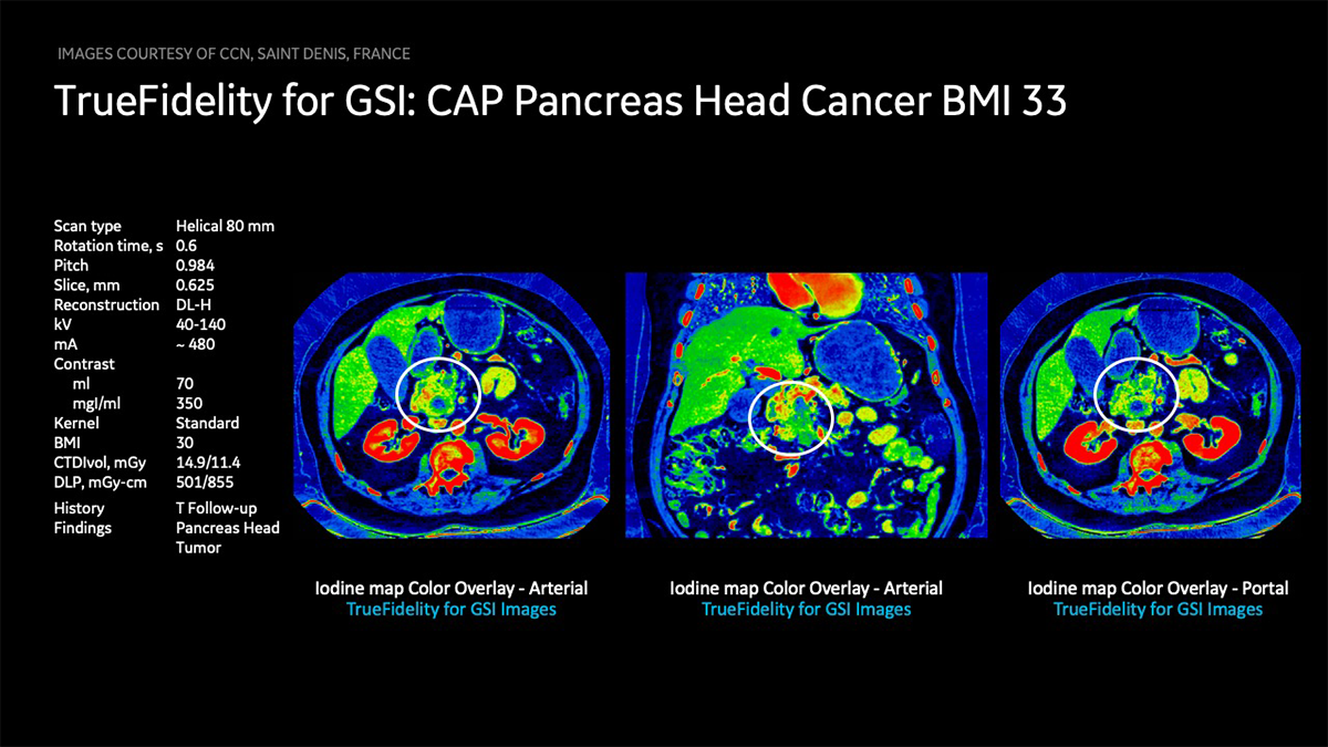
• Iodine-specific maps have the potential to increase the depiction and characterization of hypo-attenuating malignancies by increasing the contrast between a hypo-attenuating lesion and normally enhancing parenchyma on the basis of differences in tissue iodine content. Iodine-specific maps depict and quantify iodine in each voxel; thus, a small amount of enhancement in a lesion may be detected.1


_.png)


