
Senographe Pristina™
Choose the mammography system 83% of
patients say was a better experience.

Choose the mammography system 83% of
patients say was a better experience.

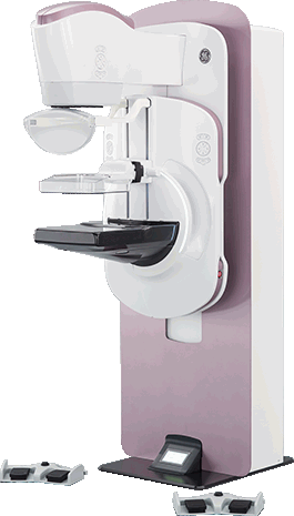
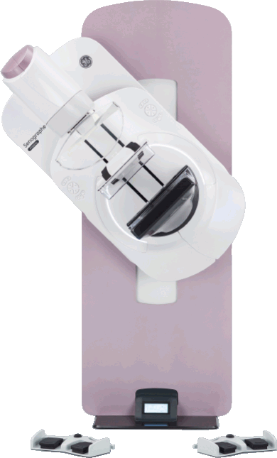
Every woman is unique. Design can make a difference to better deal with challenging patients. The new, inviting gantry with elegant lighting and gentle, rounded shapes promotes a sense of calm.
Poor positioning has been found to be the cause of most clinical imagedeficiencies and most failures of accreditation1. Technologist comfort is intimately linked to the patient’s one. The ergonomic design of Pristina eases patient positioning and improves the overall mammography experience for both patients and technologists.
Superior diagnostic accuracy2 at the lowest patient dose of all FDAapproved digital breast tomosynthesis (DBT) systems3.
Senographe Pristina sets the bar high for diagnostic confidence and performance, leveraging the Senographe family’s widely recognized image quality. The Senographe Pristina platform is upgradeable to advanced applications.

Had a more positive experience on Senographe Pristina compared to previous exams4

Of technologists report thatSenographe Pristinawas easy to use
Digital
Mammography
Digital Breast
Tomosynthesis
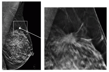
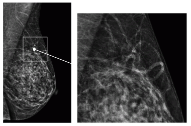
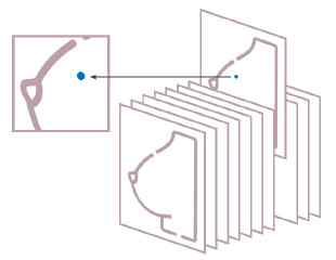
Digital Breast Tomosynthesis (DBT) is a three-dimensional (3D) imaging technology that uses a low-dose limited angle X-ray sweep around compressed breasts to produce 3D images that reduce image quality issues associated with overlapping structures (superimposition), a limitation in standard two-dimensional (2D) mammography.
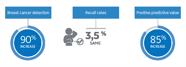
Breast cancer detection randomized trial on tomosynthesis -Reggio Emilia5: 2D + 3D mammography for finding breast cancer compared to 2D mammography alone
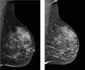
FFDM (left) and V-Preview 4.1 (right) for same patient
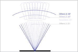
9 projections over 25° has been chosen to preserve the visibility of small objects and tissue depth separation.
Senographe Pristina mammography system is more inviting and more comfortable resulting in a better overall breast exam experience. It was designed to ease anxiety when the patient enters the exam room.
Reading tomosynthesis cases means being able to accurately interpret extensive amounts of data.
Deep learning based ProFound™ AI is changing the paradigm by offering radiologists the possibility to address the challenges of reading an increasing amount of tomosynthesis cases with a high-performing, concurrent-read, workflow solution that rapidly and accurately analyzes each tomosynthesis plane, detecting both malignant soft tissue densities and calcifications with exceptional accuracy.
Reduce
reading time
Increase
sensitivity
Increase
specificity
Reduce
recalls
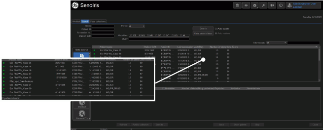
Seamless integration with Seno™ Iris: ProFound AI displays a case scores in the worklist
ProFound AI detect both malignant soft tissue densities and calcifications with exceptional accuracy findings are marked on the 2D synthetized view, on the slabs and on the planes of interest