Discover revolutionary patient care in your Breast Department with cutting-edge technologies:
LOGIQ™ Ultrasound, Invenia™ Automated Breast Ultrasound and Pristina Via™ Mammography.
Discover revolutionary patient care in your Breast Department with cutting-edge technologies that supplement one another to deliver revolutionary
patient care: LOGIQ Ultrasound, Invenia Automated Breast Ultrasound and Pristina Via Mammography.
These three cutting-edge technologies work together, enhancing diagnostic confidence, workflow efficiency, and patient comfort.
KOIOS is the first and only patented, proprietary, clinical decision support (DS) software using AI and machine learning to help to categorize the lesion. Clinically proven to improve patient outcomes and streamline workflow.
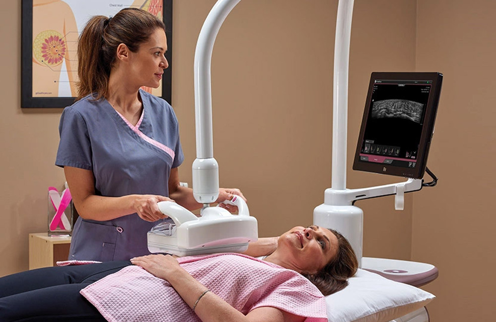
The automated ultrasound examination of the breast with Invenia Automated Breast Ultrasound offers an increase in sensitivity when finding invasive breast cancer in the dense breast tissue if Invenia Automated Breast Ultrasound is used in addition to mammography.
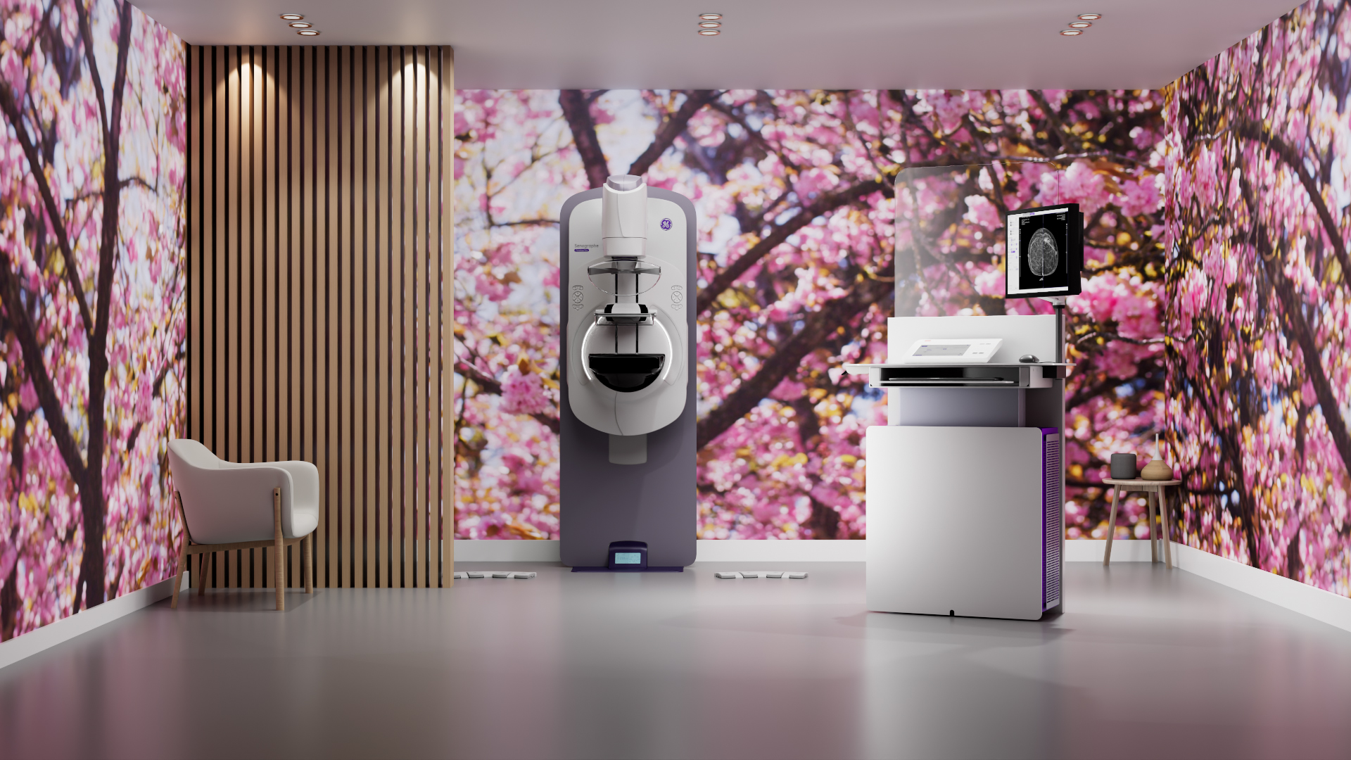
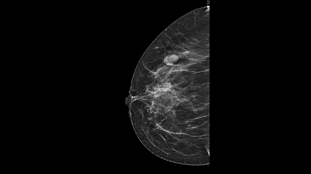
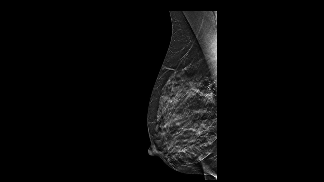
The reliable platform sets a new standard in advanced mammography—built for ease and built for you.

Regular mammography screening exams can reduce breast cancer mortality for women between the ages of 50-69 years by up to 35%1,2,3. Adding Invenia Automated Breast Ultrasound to mammography screening can increase sensitivity by 57% when detecting breast cancer for women with dense breasts.
Improved patient experience
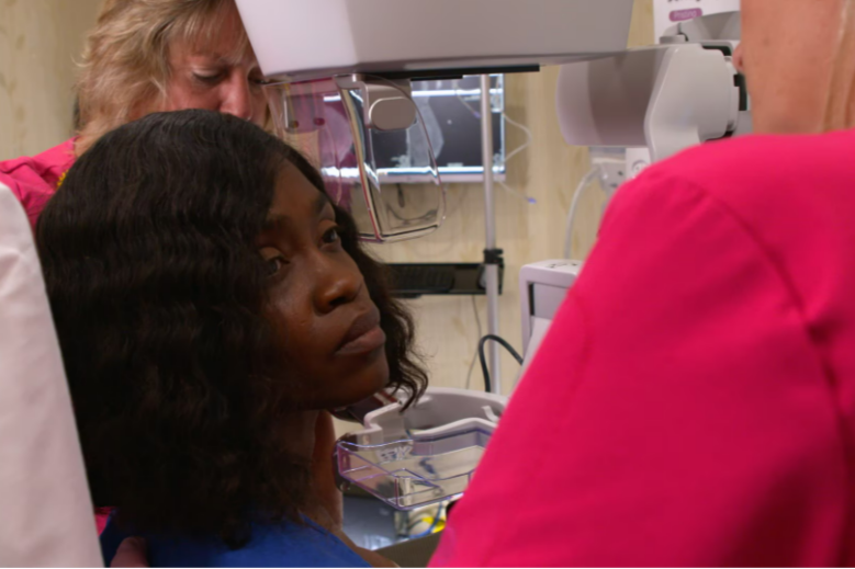
Senographe Pristina was built with one objective in mind to ease patient anxiety when they enter the exam room. This has positive effect on the mammography experience for the woman, but also makes the exam easier and more ergonomic to perform for the staff which in the end improves the quality of exams.
Invenia Automated Breast Ultrasound examination with the reverse curve transducer and the selectable compression levels reduce the patient pain*.
Faster and automated patient examination
Pristina Via offers Effortless Workflow for efficient screening examinations, with zero click exams and no waiting time for the operator. By separating the acquisition and the interpretation of the Invenia Automated Breast Ultrasound volume, the radiologist can read the entire exam in 3 minutes.
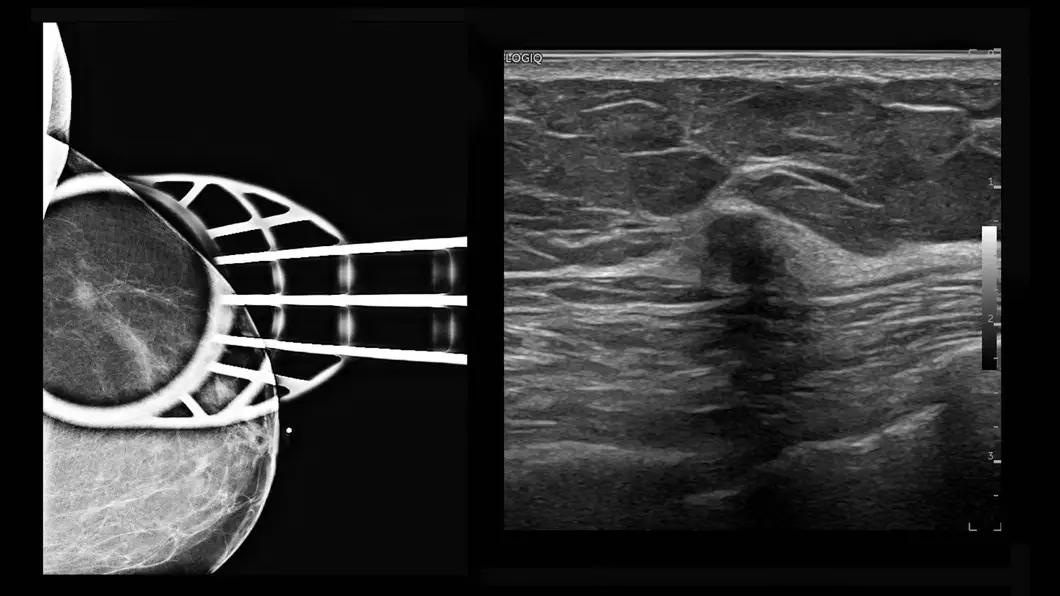
Pristina Via with Serena provides clinical confidence for cancer detection and a comprehensive solution for fast and accurate biopsies.
Invenia Automated Breast Ultrasound allows repeatable examinations in 3D and standardized bilateral evaluations. AI on LOGIQ Ultrasound systems with semi-automated segmentation and measure assistant reduces the variability of lesions categorization.
Improved accuracy with excellent image quality
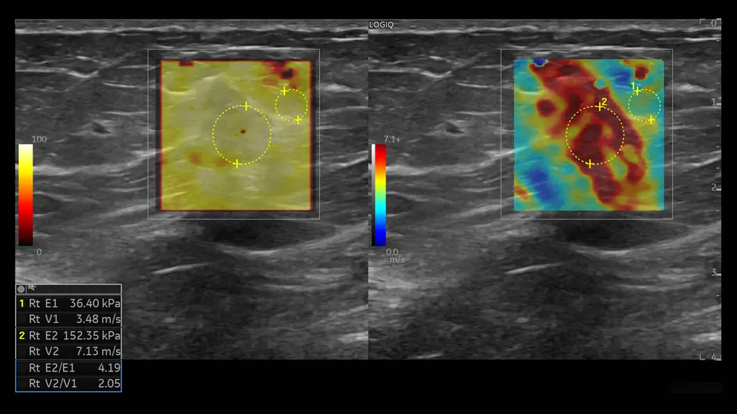
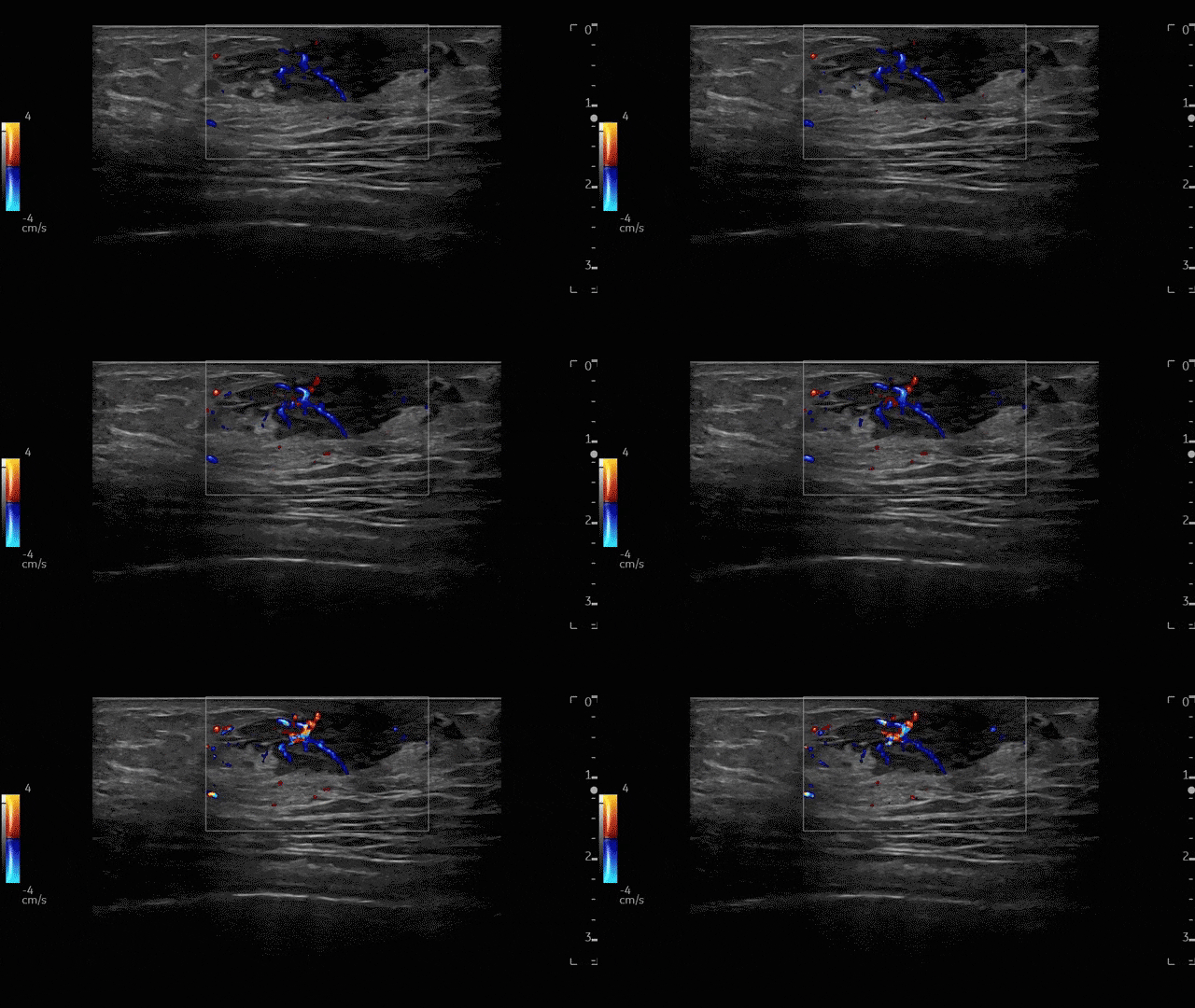
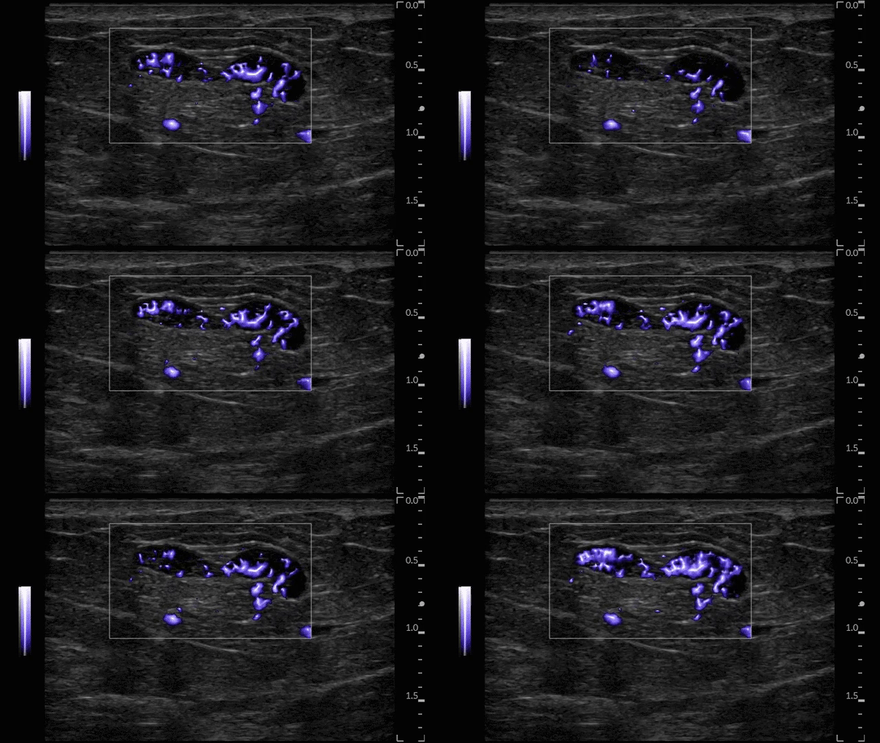
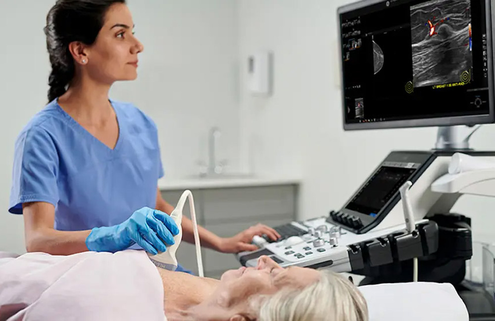
cSound Image former focusing from near field to far field enables a clear depiction and analysis of the smallest tissues abnormalities. Microvascular Imaging (MVI) High-definition flow mode and 2D Shear Wave Elastography will help you with non-invasive assessment of lesion with confident measurements.
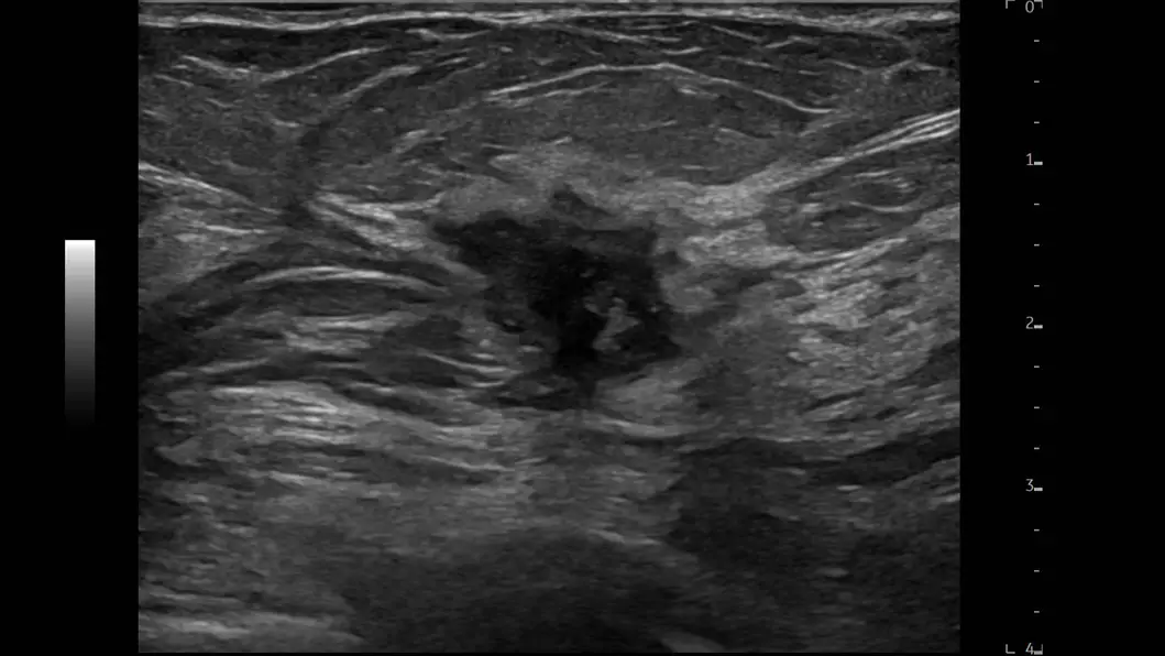
Invenia Automated Breast Ultrasound provides a full view of the breast depicting the extent of the lesion for a better surgical approach.
SenoBright HD Contrast-Enhanced Mammography reduces the masking effect of breast tissue and provides an alternative imaging method to MRI. Serena Bright CEM guided biopsy helps shorten the time frame to biopsy and diagnosis for definitive answers.
2D Shear Wave Elastography on LOGIQ’s enables non-invasive assessment of lesion stiffness in breast.
Invenia Automated Breast Ultrasound speeds up the workflow and helps with easy side-by-side comparison.
Contrast-Enhanced Ultrasound provides clear picture of tissue structure when there is a need for additional diagnosis.
The new Pristina image chain also helps reduce the exposure time in 2D with no compromise on image quality at low dose. Measurements taken on two Senographe Pristina systems consistently showed close to 30% exposure time reduction.
Non-invasive patient comfort
Invenia Automated Breast Ultrasound provides safe, non-radiation examination and reduces the cost of examination.
Breast Imaging
Exceeding your expectations
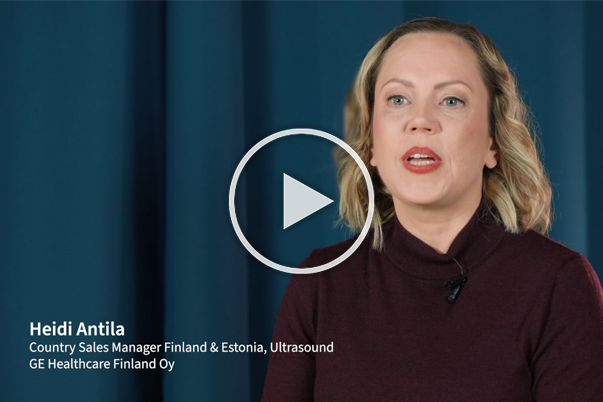
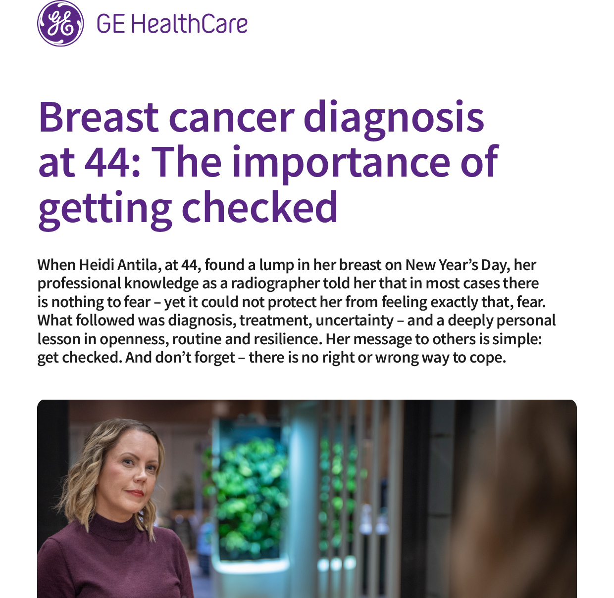
Behind every breast cancer diagnosis is a unique personal journey. Hear directly from a woman who experienced the uncertainty of diagnosis, treatment, and recovery, and learn why awareness, access to screening, and early detection can make a meaningful difference.
Hear Heidi Antila share her personal experience of discovering breast cancer, navigating diagnosis and treatment, and why early detection matters.
Read Heidi's story about uncertainty, resilience, treatment, and the lessons she learned through her breast cancer journey.
Finland, October 29-30, 2026
Sweden, September 14-17, 2027
References
*ABUS disclaimer - Shah et.al.JournalofDiagnosticMedicalSonographyDOI: 10.1177/8756479313476920 2013
1. Tabar L, Vitak B, Chen HH, Duffy SW, Yen MF, Chiang CF, Krusemo UB, Tot T, Smith RA. The Swedish Two-County Trial twenty years later. Updated mortality results and new insights from long-term follow-up. Radiol Clin North Am. 2000;38:625–651.
2. Nystrom L, Andersson I, Bjurstam N, Frisell J, Nordenskjold B, Rutqvist LE. Long-term effects of mammography screening: updated overview of the Swedish randomised trials. Lancet. 2002;359:909–919.
3. Bjurstam N, Bjorneld L, Warwick J, Sala E, Duffy SW, Nystrom L, Walker N, Cahlin E, Eriksson O, Hafstrom LO, Lingaas H, Mattsson J, Persson S, Rudenstam CM, Salander H, Save-Soderbergh J, Wahlin T. The Gothenburg Breast Screening Trial. Cancer. 2003;97:2387–2396.