

Seeing Endometriosis differently through Ultrasound
Endometriosis is a condition where tissue similar to the womb lining grows outside the uterus, often on the ovaries, fallopian tubes, or pelvic tissue, and occasionally beyond the pelvis. Its exact cause is unknown, though factors like high estrogen levels, early menstruation, heavy periods, or reproductive tract issues may increase risk. This misplaced tissue still behaves like normal endometrium—thickening, breaking down, and bleeding—but with nowhere to drain. As a result, it can become trapped, causing inflammation, scarring, cysts, and adhesions.
Without treatment, endometriosis can significantly affect fertility and may require interventions such as surgery to relieve symptoms or improve the chances of pregnancy.
Understanding Endometriosis – What, Why and How
The role of Ultrasound in Endometriosis assessment
When applied skillfully and systematically, transvaginal ultrasound goes far beyond identifying endometriomas—it can assess organ mobility, detect deep infiltrating disease, and highlight structural changes that correlate closely with symptom severity.[1][2]
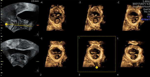
Recognising Pelvic Adhesions & Restricted Organ Mobility
Dynamic ultrasound assessment allows clinicians to evaluate organ mobility in real time, helping to identify adhesions and subtle anatomical restrictions often missed on static imaging.
Read more
Dynamic assessment of pelvic mobility is an important part of advanced endometriosis sonography. Using gentle probe pressure to test uterine and adnexal mobility helps identify loss of sliding, suggestive of adhesions [3].
The sliding sign — the absence or presence of motion between the posterior uterus/cervix/vagina and adjacent bowel — has >90% accuracy in predicting Pouch of Douglas (POD) obliteration in suspected endometriosis [4]. A major meta-analysis found strong diagnostic performance for both POD obliteration and bowel involvement [5].
Using the sliding sign also improves pre‑operative assessment, with significantly higher sensitivity compared to bimanual examination alone [6].
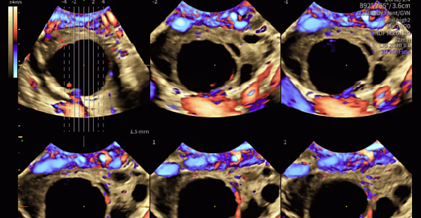
Identifying Ovarian Endometriomas through Ultrasound
Ultrasound provides clear visualisation of ovarian endometriomas, making it one of the most reliable and commonly recognised routes for detecting cystic manifestations of endometriosis.
Read more
Ovarian endometriomas demonstrate characteristic ultrasound appearances such as ground‑glass echogenicity, lack of internal vascularity, and uni‑ or multilocular cystic structures — widely described in practical pelvic ultrasound guides [3]. NICE‑aligned summaries also highlight endometriomas as a common direct sonographic sign of disease [7].
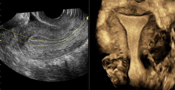
Detecting Deep Infiltrating Endometriosis (DIE)
A targeted ultrasound approach detects deep infiltrating endometriosis, revealing lesions across pelvic compartments for surgical planning.
Read more
A focused, methodical TVUS examination enables detection of DIE in structures including the uterosacral ligaments, rectovaginal septum, rectosigmoid bowel, bladder and ureters [2][8].
Findings may include echogenic fibrosis, hypoechoic nodules, kissing ovaries, and fixed retroverted uterus, all of which are strongly associated with posterior compartment disease [3][8].
The IDEA framework formalises these targets into a structured four‑step approach to ensure systematic mapping of disease severity and location [9].

Why structured scanning protocols matter in Endometriosis assessment
Using a structured, standardised protocol improves consistency, reduces diagnostic variability, and ensures thorough assessment of key endometriosis markers.
Read more
Consensus statements emphasise that structured protocols significantly improve scanning consistency, documentation, and diagnostic confidence [9].
The Society of Radiologists in Ultrasound (SRU) Consensus 2024 specifically recommends incorporating:
- posterior compartment assessment,
- uterine/ovarian positional relationships,
- dynamic mobility testing, and
- the sliding sign into routine pelvic ultrasound for at‑risk patients, noting these additions require ≤5 minutes and can help reduce diagnostic delays [1].

Blog: New Study Aims to Revolutionise the Diagnosis of Endometriosis
One in ten women of reproductive age suffer from endometriosis. Out of these women, 30-50% will experience fertility problems because of...

Free Download: Ultrasound in gynaecology imaging poster
Ultrasound has become an essential tool in the evaluation of pelvic anatomy.
Diagnosing Endometriosis with Ultrasound
Ultrasound plays a central role in the diagnostic pathway for suspected endometriosis, offering real‑time visual assessment of pelvic structures and dynamic functional testing that cannot be achieved through examination alone. When performed systematically, TVUS can identify both direct and indirect signs—supporting earlier recognition, improved triage, and more informed decision‑making across the patient journey.

Blog: How to detect Endometriosis using 3D Ultrasound
Transvaginal 3D ultrasound is a valuable but often underutilised tool in gynaecology. Research has shown it is sensitive and specific for detecting endometriosis beyond the ovaries, according to a review in the Journal of Ultrasound in Medicine.
Although an MRI is commonly ordered to detect deep infiltrating endometriosis, ultrasound has comparable sensitivity and specificity for lesions in the rectum and lower sigmoid colon, at a lower cost to boot. Ultrasound is also a more comfortable procedure for many women and can eliminate the need to travel for additional appointments...

On-demand Webinar: How to Diagnose Deep Endometriosis Using Pelvic Ultrasound
Hear from Dr. Susanne Johnson on how you can diagnose deep Endometriosis using Pelvic Ultrasound as she tackles some of the most common myths around Ultrasound diagnosis and provides insights on the systematic process behind diagnosing with Pelvic Ultrasound.
Free downloadable posters
Explore our selection of educational posters to help you navigate all aspects of Endometriosis diagnosis using Ultrasound:

The Pelvic Pain Patient
Easy 5-Step Ultrasound Evaluation

Ultrasound assessment of
deep infiltrating endometriosis

Ultrasound and MRI scanning
For management of endometriosis

Classification of
uterine anomalies

Ultrasound Imaging
of Uterine Malformations

Systematic approach to sonographic
evaluation of the pelvis

The Endometrium on Ultrasound
Endometrial Assessment

Ultrasound in
gynaecology imaging
Why early imaging matters
Symptoms of endometriosis overlap with many benign conditions, making imaging essential even when examinations are normal. NICE guidance stresses that a normal TVUS does not exclude endometriosis when clinical suspicion remains high [7].

Earlier Clarity in a Complex Condition
Symptoms of endometriosis often mimic other pelvic or gastrointestinal conditions, making early imaging key to differentiating causes. NICE highlights that even a normal ultrasound doesn’t rule out endometriosis when symptoms point strongly toward the diagnosis[7].

Reducing Long Diagnostic Delays
People in the UK currently face an average diagnostic delay of almost nine years, contributing to worsening symptoms, disease progression and reduced quality of life[10]. Early imaging helps shorten this pathway by identifying specific ultrasound markers linked to endometriosis[11].

Improved Referral and Care Pathway Planning
Detecting features such as endometriomas, adhesions or reduced pelvic mobility can guide clinicians toward earlier specialist referral and more appropriate management decisions.

Anticipating Surgical Complexity
Ultrasound findings—particularly a negative sliding sign indicating potential pouch of Douglas obliteration—help predict more complex surgery and allow teams to plan accordingly.[6]

“For six months I really couldn’t do anything. I could barely function. It was really scary because I wondered if this was going to be my life now?”
Women with endometriosis wait an average of 7.5 years before they receive a proper diagnosis. Follow a mother & daughter’s search for answers about pelvic pain.
Frequently Asked Questions (FAQs): Endometriosis diagnosis, facts and treatment
Here are some common questions from physicians making an endometriosis diagnosis or treatment plan.
What do you need to know about endometriosis diagnosis and treatment?
As a gynaecologist, it's important to recognise the link between infertility and endometriosis. When making an endometriosis diagnosis or treatment plan, using 3D ultrasound can help ensure early and accurate detection as well as map out treatment options. Read more
What should your patients understand about endometriosis?
Make sure your patients know the symptoms of the disorder, including gastrointestinal and urinary tract symptoms. A history of dysmenorrhea can also indicate endometriosis. Read more
Can endometriosis cause endometrial cancer?
Endometriosis may lead to a greater risk of endometrial cancer. For endometrial cancer, ultrasound is indeed the key to early detection. While classic symptoms and signs are well described in the literature, a point-of-care ultrasound can help measure endometrial thickness and detect endometrial cancer in its early stages. Read more
Is endometriosis genetic?
Polygenic inheritance, or more than one gene controlling a disease, is thought to be responsible for endometriosis, but the exact cause is not certain. Learn how to discuss the genetics of endometriosis with your patients. Read more
Should you diagnose endometriosis with MRI or transvaginal ultrasound?
Diagnosing endometriosis is a complex process that often includes the use of both ultrasound and MRI. Comparing the results of these two imaging modalities, including using 3D ultrasound when possible, can assist in diagnosing endometriosis. Read more
How can you detect endometriosis using 3D ultrasound?
As a gynaecologist, knowing how to detect endometriosis with 3D ultrasound is an important skill. While laparoscopy is the gold standard, 3D ultrasound can add important information before surgery, particularly when evaluating deep infiltrating endometriosis. Read more
Additional resources

Voluson Club
The only ultrasound community dedicated to the education and collaboration of women’s health practitioners to help expand your expertise and your practice.

Voluson Education
Your dedicated portal for clinical resources and product information related to women’s health, including a clinical posters library and educational content.
Want to learn more about our solutions for Endometriosis diagnosis?
Privacy Policy | Terms And Conditions | Contact Us | Communication Preferences | Unsubscribe
© 2026 GE HealthCare. Voluson is a trademark of GE HealthCare. GE is a trademark of General Electric Company used under trademark license. JB00557IE.
Commercial availability of GE HealthCare medical systems is subject to meeting local requirements in a given country or region. Contact a GE HealthCare Representative for more information. Intended for healthcare professionals only.
[1] Society of Radiologists in Ultrasound (SRU). Consensus on Routine Pelvic US for Endometriosis. Radiology. Apr 9, 2024. [2] Jing Fang. Ultrasound Assessment of Deep Infiltrating Endometriosis. Monash Health. [3] BMUS. Ultrasound for Endometriosis – Quick Reference Guide. [4] Radiopaedia. Sliding Sign – Sonographic Interpretation. [5] Alcázar JL, et al. Diagnostic Accuracy of the Sliding Sign… Ultrasound Obstet Gynecol. 2022;60(4):477–486. [6] Arion K, et al. Prediction of POD Obliteration: TVUS vs Pelvic Exam. J Minim Invasive Gynecol. 2019. [7] NICE. Endometriosis: diagnosis and management (NG73). Updated 2024. [8] Clinical DIE mapping findings: Monash Health (Jing Fang Workshop). [9] ISUOG / IDEA Group. Systematic Sonographic Evaluation of Endometriosis. [10] Endometriosis UK. Diagnosis Survey 2023 Report. [11] Frontiers in Medicine. Systematic Review on Factors Contributing to Delayed Diagnosis. 2025.